

The infant suffered from severe hydronephrosis due to obstruction, adhesion, and a high insertion of the ureter. The patient was placed in an oblique supine position. An extraperitoneal flank incision was made guided by the location of the 11th rib.

After opening the Gerota's fascia, a vessel loop was used for easy manipulation of the ureter during surgery.

The ureter was liberated from adhesion bands found throughout the perirenal space, potentially attributed to recurrent infections.

In figure 5, a segment of the ureteropelvic junction was removed with incisions made on both the renal pelvis and ureter. A diamond-shaped cut is made on the renal pelvis and a T-cut on the ureter side, which includes an oblique-transverse and a longitudinal cut.
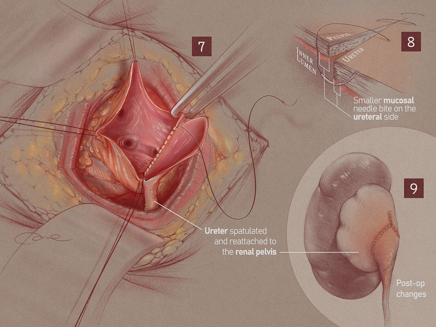
The ureter was then spatulated and reattached to the renal pelvis as shown in figure 7. Figure 8 shows that there should be smaller mucosal needle bites taken on the ureter side, to prevent future ureteral obstruction from the sutures.

This is the pre- and post-op changes in the ureteropelvic junction.


